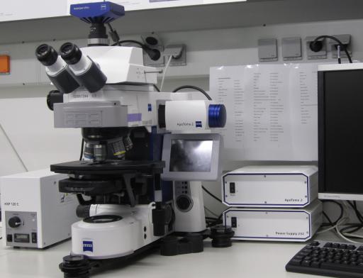
Structured Illumination Microscope (Zeiss ApoTome - DZNE W1) Zeiss Axio Imager.M2
Basic Information
| Name: | Structured Illumination Microscope (Zeiss ApoTome - DZNE W1) | |
| Manufacturer: | Zeiss | |
| Model: | Axio Imager.M2 | |
| Facility: | Light Microscopy Facility (LMF) | |
| Partner: | German Center for Neurodegenerative Diseases Within The Helmholtz Association (DZNE) | |
Description
- Category
- Light Microscope
- Overview
- It is designed for routine work on fixed samples. Optical sectioning can be obtained on fixed samples by the use of the Apotome.
- Suitable for fixed samples, fluorescently labeled samples (structured illumination possible), histologically fixed samples (no strucutred illumination), big samples
- Features
- Upright stand
- motorized XY stage (Märzhäuser SMC2009)
- motorized z-drive
- fluorescence
- transmitted light with DIC
- Darkfield b/w epifluorescence acquisition
- Optical Sectioning (ApoTome2)
- sequential imaging
- Brightfield and Fluorescence overlay
- timelapse
- z-stack
- tile scans
- multi position experiments
- complex experiments via Experiment Designer
- optical sectioning
- Objectives
- Zeiss EC Plan Neofluar 5x 0.16
- Zeiss Plan-Apochromat 10x 0.45
- Zeiss Plan-Apochromat 20x 0.8
- Zeiss Plan-Apochromat 40x 0,95 korr.
- Zeiss EC Plan-Neofluar 100x 1.3 Oil
- Illumination
- Fluorescence (Metal Halide, HXP, 120W)
- Transmitted Light (Halogen)
- Detection
- Axiocam MRm rev.3 (1388*1040
- 6,45*6,45µm)
- Reflectors
- FS 49 (DAPI) : EX BP G365
- BS 395
- EM BP 445/50
FS 38 HE (GFP) : EX BP 470/40 - BS 495
- EM BP 525/50
FS 20 (Rhodamine) : EX BP 546/12 - BS 560
- BP 575-640
FS 50 (Cy5) : EX BP 640/30 - BS 660
- EM 690/50
- FS Analysator for DIC and Transmission
- Software
- ZEN Blue 2012
- Incubation
- Not available
Link to Further Details
Points of Contact
Images
Last Update
Last updated at: 8 April 2018 at 03:11:23

 View all instruments of this unit
