 View all instruments of this unit
 Lattice Lightsheet Microscope (LLSM) Custom built
Basic Information
| Name: | Lattice Lightsheet Microscope (LLSM) | |
| Manufacturer: | Custom built | |
| Facility: | Advanced Imaging Facility (AIF) | |
| Partner: | Max Planck Institute of Molecular Cell Biology and Genetics (MPI-CBG) |
|
| Location: | 202 | |
Description
Lattice lighsheet microscope (LLSM)
Originally developed by Eric Betzig (link), this microscope relies on illuminating the sample with a coherent array of Bessel beams in combination with a high numerical aperture lens on the detection side for achieving high optical sectioning. The setup offers:
- High optical sectioning (≤ 500 nm)
- Low photodamage
- Fast acquisition (up to 300-400 FPS, single plane)
The acquired data can be deconvolved using a GPU-accelerated algorithm, available on-line during image acquisition. Best used with thin, transparent samples (adherent cells, early embryos).
Microscope configuration
Available laser lines:
- 488 nm
- 561 nm
- 589 nm
- 642 nm
Detection objectives:
- Nikon 25x/1.1 Water immersion
Illumination objective:
- Special Optics 28.6x/0.7 Water immersion (custom objective)
Imaging camera:
- Dual Hamamatsu ORCA Flash v4.0
Notes
- The microscope uses a custom sample holder
- Samples must be attached to 5 mm diam. round coverslips (available at AIF)
- Samples are in liquid environment during imaging
- Temperature/CO2 controller available
Link to Further Details
https://www.mpi-cbg.de/en/services-facilities/core-facilities/advanced-imaging-facility/microscopes/
Options of instrument usage
- This instrument can be used with the support of a supervising assistant.
Points of Contact
Access Requirements
Access rules
Basic Users can only access the instruments after electronic booking, and with an Assistant. Advanced Users might be allowed to access the instruments without supervision, but only after having received a proper training on the microscope by an Assistant. In any case, a User will always be able to contact an Assistant during normal working hours using the internal telephone system for troubleshooting unexpected issues.
Questions concerning the sample mounting and handling will be discussed before the first training session, in order to maximize the time a User will actually spend collecting data.
Images
| Preview | Description |
|---|---|
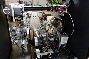
|
Lattice Lightsheet - main assembly |
Last Update
Last updated at: 19 November 2018 at 21:06:35