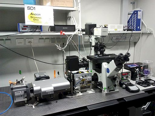
Real-Time Confocal Microscope (SD1 - Andor Spinning Disc) Olympus IX71
Basic Information
| Name: | Real-Time Confocal Microscope (SD1 - Andor Spinning Disc) | |
| Manufacturer: | Olympus | |
| Model: | IX71 | |
| Facility: | Light Microscopy (LMF) | |
| Partner: | Max Planck Institute of Molecular Cell Biology and Genetics (MPI-CBG) | |
| Inventory number: | 0 10.1.65.208 | |
Description
- Category
- Light Microscope
- Overview
- Fast, "real-time" confocal imaging. Laser equipment: 488 nm solid-state Coherent Sapphire (75mW), 561 nm solid-state Cobolt Jive (75mW) + 640 Coherent Cube (40mW) .
- Suitable for confocal imaging of EGFP, mCherry, dTomato, Draq5 and the like with high temporal and spatial resolution.
- Features
- Inverted stand
- manual
- 1.6x OptoVar
- fast piezo objective z-positioner (Physik Instrumente - PI)
- Prior ProScanIII xy scanning stage
- micro point with 15Hz cutter laser with 365nm and 405nm dye cells;
- objectives 40x 1.25 SIL and 60x 1.3 SIL are shared objectives and need to be booked separately via the LMF booking database!
- Objectives
- Olympus UPlanSApochromat 10x 0.4
- Olympus UPlanFluar 20x 0.5
- Olympus UPlanFluarN 60x 0.9
- Olympus UPlanSApochromat 60x 1.20 W
- Olympus UPlanSApochromat 100x 1.4 Oil
- Olympus UPlanSApochromat 40x 0.9
- Olympus UApochromat 40x 1.15 W
- Olympus UPlanFLN 40x 0.75 Ph2
- Olympus UPlanFluar 60x 1.25 Oil Ph3
- Olympus UApochromatN340 40x 1.15 W
- Illumination
- Transmitted Light (Halogen)
- Fluorescence (HBO)
- Laser DPSS 488 nm
- Laser DPSS 561 nm
- Laser DPSS 640 nm
- Detection
- [[detection::Spinning disc scan head Yokogawa CSU-X1 (10.000rpm):
dichromatic mirrors:
Quad band T-405/488/568/647
Triple band T-405/488/561
Single band T-488
physical pinhole radius: 25um - physical pinhole spacing: 250um
- back-projected pinhole radius: 0.15um
- back-projected pinhole spacing: 1.5um (with 100x objective
- 1.6x optovar)
Andor Dual Camera Port TuCam with two cameras and 0.95x projecting lens for each camera: - Andor iXon EM+ DU-897 BV back illuminated EMCCD
- dexel size of EMCCD chip: 16um
Andor Neo sCMOS - dexel size of CMOS chip: 6.5um
image pixel sizes (measured April 3rd 2012):
for use with iXon: with 10x air objective: 1.695um/px (1x optovar) - 1.055 um/px (1.6x opt.)
- with 40x air obj.: 0.418um/px (1x opt.)
- 0.26um/px (1.6x opt.)
- with 60x W obj.: 0.279um/px (1x opt.)
- 0.174um/px (1.6x opt.)
- with 100x oil obj.: 0.168um/px (1x opt.)
- 0.105um/px (1.6x opt.)
for use with Neo: with 10x air objective: 0.692um/px (1x optovar) - 0.432um/px (1.6x opt.)
- with 40x air obj.: 0.17um/px (1x opt.)
- 0.106um/px (1.6x opt.)
- with 60x W obj.: 0.114um/px (1x opt.)
- 0.071um/px (1.6x opt.)
- with 100x oil obj.: 0.068um/px (1x opt.)
- 0.043um/px (1.6x opt.)
old measure pixel sizes before major upgrade 26th - 30th March 2012:
image pixel size (measured again after major scope repair on March 7th 2011): with 100x objective: 0.175um (was 0.178um before 20110307) - 100x obj
- and 1.6x optovar: 0.109um (was 0.111um before 20110307)
- 60x obj.: 0.289um (was 0.288um before)
- 60x obj
- and 1.6x opt.: 0.180um (was 0.181um before 20110307)
- 10x obj. : 1.747um
- 10x obj
- and 1.6x opt.: 1.089um
camera projecting lens has about 0.92x (was 0.9x before 2011_03_07) magnification calculated from the measured pixel sizes]]
- Reflectors
- Optical Insight DualView image splitter (currently not installed):
holder for GFP/mCherry: BL 525/40 - BS 565
- ET 605/70
holder for GFP/RFP: HC 520/35 - BS 565
- HC 628/40
Rotr filter wheel:
position 0: 624/40 - 1: open
- 2: 525/30
- 3: razor edge 568LP
- 4: dual emitter: 512/23+630/91
- 5: razor edge 488LP
- 6: old CSU dual emitter: 525/30 + 650/?
- 7: 605/70
- 8: 445/40
- 9: edge basic 635LP
microscope filter turret:
position 1: GFP cube - 2: RFP cube
- 3: dual GFP + RFP cube
- 4: cube with z375 RDC for micropoint cutter
- 5: empty
- 6: DIC analyzer
- Software
- [[software::iQ 2.9 + iQ 3.0 (both with Python) iQ3 user guide]]
- Incubation
- Stage incubator (T) + objective heater
Link to Further Details
Points of Contact
Images
Last Update
Last updated at: 8 April 2018 at 03:11:05

 View all instruments of this unit

